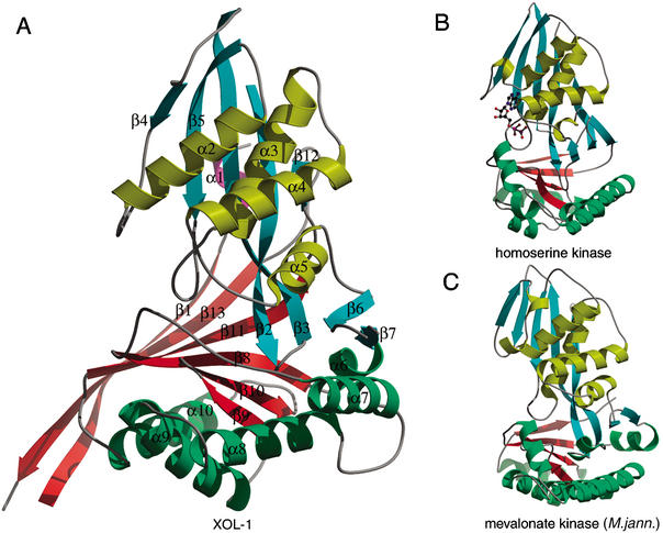
Figure 2.
Comparison of XOL-1 and GHMP kinase structures. (A) The structure of XOL-1. Ribbon diagram of XOL-1 (PDB ID: 1MG7). Domain 1 consists of β-strands 2–7 and 12 (cyan) and α-helices 1–5 (yellow). Domain 2 consists of β-strands 8–11 (red) and α-helices 6–10 (green). Additional strands β1 and β13 and helix α1 form novel elements not found in known GHMP kinase structures. (B) The structure of homoserine kinase bound to ADP (PDB ID: 1FWK). Domain 1 β-strands and α-helices are cyan and yellow, respectively. Domain 2 β-strands and α-helices are red and green, respectively. ADP is represented by a ball and stick model. (C) The structure of mevalonate kinase (M. jannaschii; PDB ID: 1KKH). Colors are as in B.

