Figure 5.
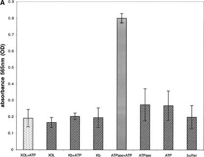
XOL-1 ATP hydrolysis and binding. (A) In the presence of XOL-1 (10 μg; stippled bar), absorbance at 565 nm (Y-axis) is not significantly increased over the absorbance observed in any negative controls (hatched bars) in an enzyme-coupled colorimetric ATPase assay (see Materials and Methods). Negative controls (hatched bars) XOL-1, H-2Kb + 1 mM ATP, H-2Kb, Na+/K−-ATPase (5 milliunits), 1 mM ATP, buffer. As a positive control (gray bar), N+/K−-ATPase (5 milliunits) was incubated with 1 mM ATP. (B) The fluorescence emission of 20 mM ATP-TNP (○) is not significantly increased in the presence of 20 μM XOL-1 (▴). As a positive control, 20 μM hexokinase was incubated with 20 μM TNP-ATP (▵). Fluorescence emission intensity (Y-axis) is represented as function of wavelength (505–600 nm, X-axis). Excitation wavelength = 410 nm.


