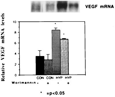
Figure 5.
Effect of wortmannin on hypoxic induction of VEGF. (Upper) Northern blot. From left to right, the first lane represents mRNA from SVR cells under normoxic conditions in the absence of wortmannin. The second lane represents SVR cells under normoxic conditions in the presence of 1 μg/ml wortmannin. The third lane represents mRNA from SVR cells under hypoxic conditions in the absence of wortmannin, and the fourth lane represents mRNA from SVR cells under hypoxic conditions in the presence of wortmannin. (Lower) Normalized mRNA levels, labeled as in Fig. 4.

