Figure 3.
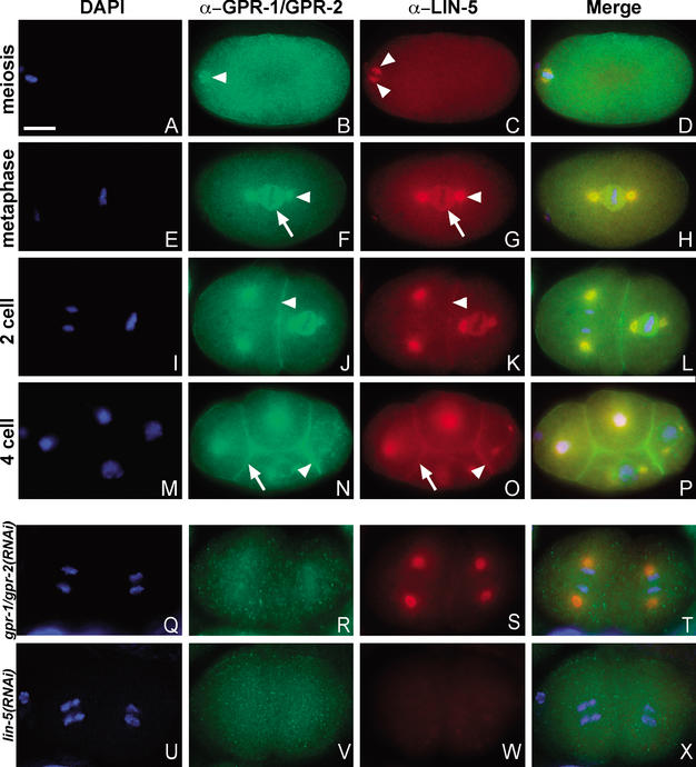
GPR-1/GPR-2 localize to the mitotic spindle and cell cortex in a LIN-5-dependent manner. DNA staining with DAPI (blue) and immunostaining of GPR-1/GPR-2 (green) and LIN-5 (red). Merged images are shown in D, H, L, P, T, and X. (A–D) Fertilized embryo in meiosis II. LIN-5, but not GPR-1/GPR-2, showed specific localization to the meiotic spindle (arrowheads, B,C). (E–H) One-cell embryo in metaphase. GPR-1/GPR-2 (F) and LIN-5 (G) colocalize to the spindle asters (arrowheads) and kinetochore MTs (arrows). (I–L) Two-cell embryo. GPR-1/GPR-2 (J) and LIN-5 (K) colocalize to the spindle apparatus as well as the cell cortex (arrowheads). (M–P) Four-cell embryo. In addition to spindle staining, both proteins showed stronger staining at the P2/EMS boundary (arrowhead) than at other cell membranes (arrow). (Q–X) Localization in RNAi embryos. (Q–T) GPR-1/GPR-2 staining is strongly reduced but not fully eliminated in gpr-1/gpr-2(RNAi) embryos. (S) LIN-5 staining remained at the spindle apparatus but appeared reduced at the cell periphery. (U–X) lin-5 RNAi eliminated LIN-5 staining (W) and disrupted GPR-1/GPR-2 localization (V). Bar, 10 μm.

