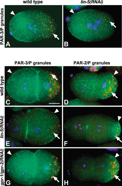
Figure 4.
gpr-1/gpr-2 and lin-5 do not determine polarity in the early embryo. Embryos are shown with P granules in red (arrows), DNA in blue (DAPI), and either PAR-3 (A–C,E,G) or PAR-2 (D,F,H) in green. (A,B) lin-5 is not required to establish polarity in the one-cell embryo. PAR-3 (arrowheads) localizes to the anterior cortex in wild-type (A) and in lin-5(RNAi) (B) one-cell embryos, and P granules localize to the posterior in both embryos. (C,D) Two-cell wild-type embryos. PAR-3 localizes to the entire cortex of the anterior AB cell and the anterior region of the posterior P1 cell (C, arrowhead). PAR-2 (D, arrowhead) and P granules (arrows) localize to the posterior in P1. (E,F) Two lin-5(RNAi) embryos in which AB is smaller than P1. PAR-3 (E, arrowhead) localized to the P1 cortex and posterior of AB. PAR-2 (F, arrowhead) localized to the posterior cortex of P1, although at times in a more restricted area. P granules show wild-type localization. The punctate PAR staining at the cortex between the blastomeres is not reproducible. (G,H) Two gpr-1/gpr-2(RNAi) embryos with AB and P1 of similar size. PAR-3 (G, arrowhead), PAR-2 (H, arrowhead), and P granules (arrows) localized like in wild type. Bar, 10 μm.

