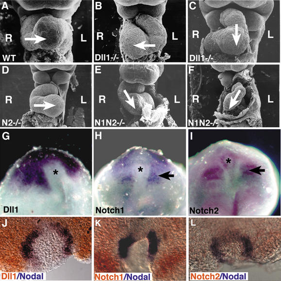
Figure 1.
Heart looping defects in Notch pathway mutants. (A–F) Scanning electron micrographs of embryos at E9.5. The direction of heart looping is indicated by the arrow. The left (L) and right (R) sides of the embryo are indicated. (A) Wild-type embryo exhibiting normal right-to-left heart looping. (B) Dll1−/− mutant embryo exhibiting reversed heart looping. (C) Dll1−/− mutant embryo exhibiting ventral heart looping. (D) Embryo homozygous for the Notch2del1 hypomorphic allele (designated N2−/−) exhibiting normal right-to-left heart looping. (E) Notch1−/− Notch2−/− double-mutant embryo exhibiting mild ventral looping. (F) Severely affected Notch1−/− Notch2−/− double-mutant embryo exhibiting reversed looping and cardiac hypoplasia. (G–I) Expression of Notch pathway genes around the node (asterisk). (G) Dll1 expression. (H) Notch1 expression. (I) Notch2 expression. Both Notch1 and Notch2 are also expressed in the paraxial mesoderm (arrows). (J–L) Double-label whole-mount in situ hybridization with a digoxigenin-labeled riboprobe (blue) for Nodal and fluorescein-labeled riboprobes (orange) for Dll1 (J), Notch1 (K), or Notch2 (L).

