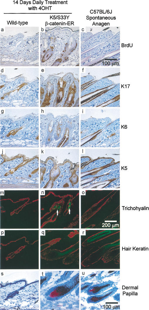
Figure 3.
Altered expression of hair follicle markers in K5/S33Yβ-catenin–ER mice. Immunohistochemistry and immunofluorescence were performed on parasagittal sections of skin from both wild-type and K5/S33Yβ-catenin–ER mice in addition to a normal, spontaneous anagen cycle in a C57BL/6J mouse. The markers used were as follows: BrdU (a–c), K17 (d–f), K6 (g–i), K5 (j–l), trichohyalin (m–o), hair keratin (p–r), and alkaline phosphatase staining for dermal papillae (s–u). White arrows highlight ectopic trichohyalin staining in n. Bars: a–l,s–u, 100 μm; m–r, 200 μm.

