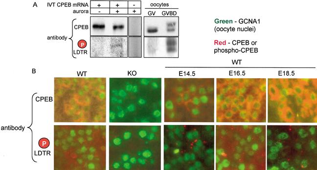
Figure 2.
CPEB phosphorylation is regulated during prophase I progression. (A) CPEB mRNA was translated in a reticulocyte lysate, some of which was then supplemented with recombinant aurora. The proteins were then Western blotted and probed with general CPEB antibody or phospho-specific antibody. Some lysate that was not primed with any RNA was supplemented with aurora and then probed with both CPEB antibodies. GV and GVBD oocytes were also probed with the general and phospho-specific CPEB antibodies. Although only nonphosphorylated CPEB is present in GV stage oocytes (Tay et al. 2000; Hodgman et al. 2001), aurora-phosphorylated CPEB is present after GVBD. Some CPEB is additionally phosphorylated by cdc2 at this time, which slows its electrophoretic mobility (Mendez et al. 2000; Tay et al. 2000). (B) Ovaries from E16.5 wild-type and CPEB knockout animals were fixed, embedded, sectioned, and immunostained with general CPEB antibody or the phospho-specific antibody. The sections were also immunostained for GCNA1, a marker for oocyte nuclei. Note that although both the general and phospho-specific antibodies were immunoreactive with the wild-type ovaries, there was no signal with the knockout ovaries. Ovaries from wild-type E14.5, E16.5, and E18.5 ovaries were immunostained with the same three antibodies noted above. Although general CPEB immunoreactivity was detected at all three stages, the phospho-specific antibody was immunoreactive only with the E16.5 ovary section.

