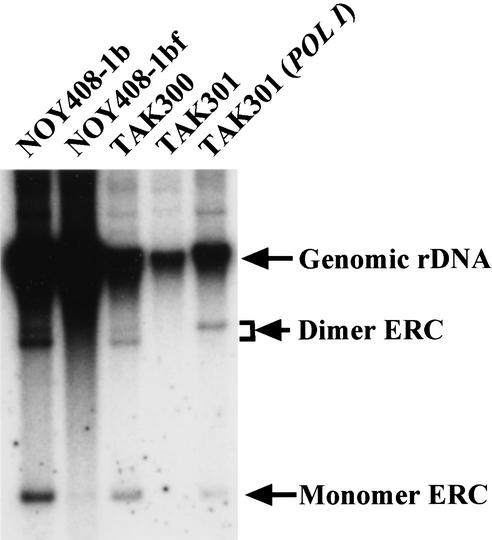
Figure 3.
Detection of the extrachromosomal rDNA circles (ERCs). DNA isolated from various strains was subjected to electrophoresis (0.6% agarose gel for 20 h at 1 V/cm) followed by Southern hybridization using the rDNA probe (see Fig. 1). The positions of monomer ERC, dimer ERC, and genomic rDNA are indicated by arrows. The strains are the same as used in Figure 2C. We used the same number of cells, which were determined by spectrometer to isolate the DNA.

