
Figure 3. Expression analysis of TRP channels in the murine organ of Corti.
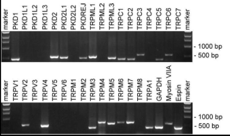
As a template, cDNA from an organ of Corti library (a gift from Dr. Bechara Kachar, NIH, Bethesda, USA) was used to PCR amplify TRP channel fragments with specific sense and antisense primers. Primers that did not give signals in this analysis were functionally verified with cDNA from other mouse tissues (not shown).