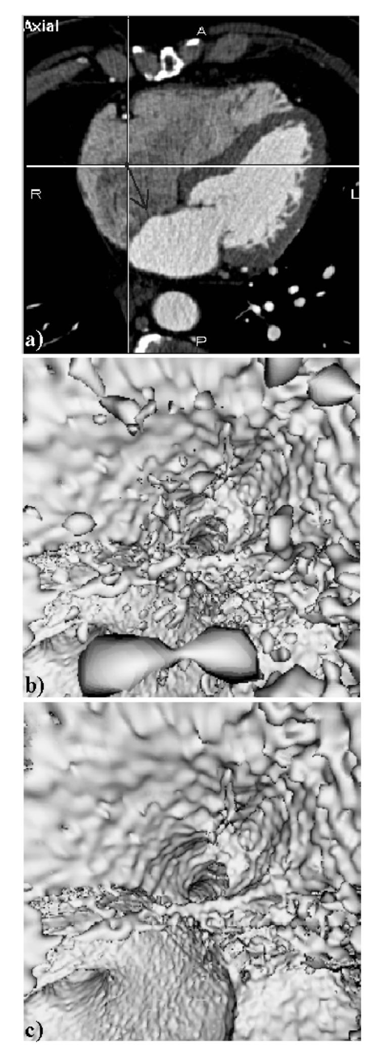
Fig. 3.

Inside the right atrium, (a) Axial MSCT slice showing the location and the orientation of the virtual sensor (dark arrow) located in the right atrium and looking towards the entrance of the coronary sinus, (b) Virtual endoscopy image computed without region growing (CT = 70 HU). The artifact regions to be eliminated appear like isolated particles in suspension within the atrium cavity, (c) Virtual endoscopy image computed with region growing (CT = 70 HU, ST = 1500 voxels). The entrance of the coronary sinus (at the centre of the image) and the surrounding endocardial wall (top half of the image) are clearly visible.
