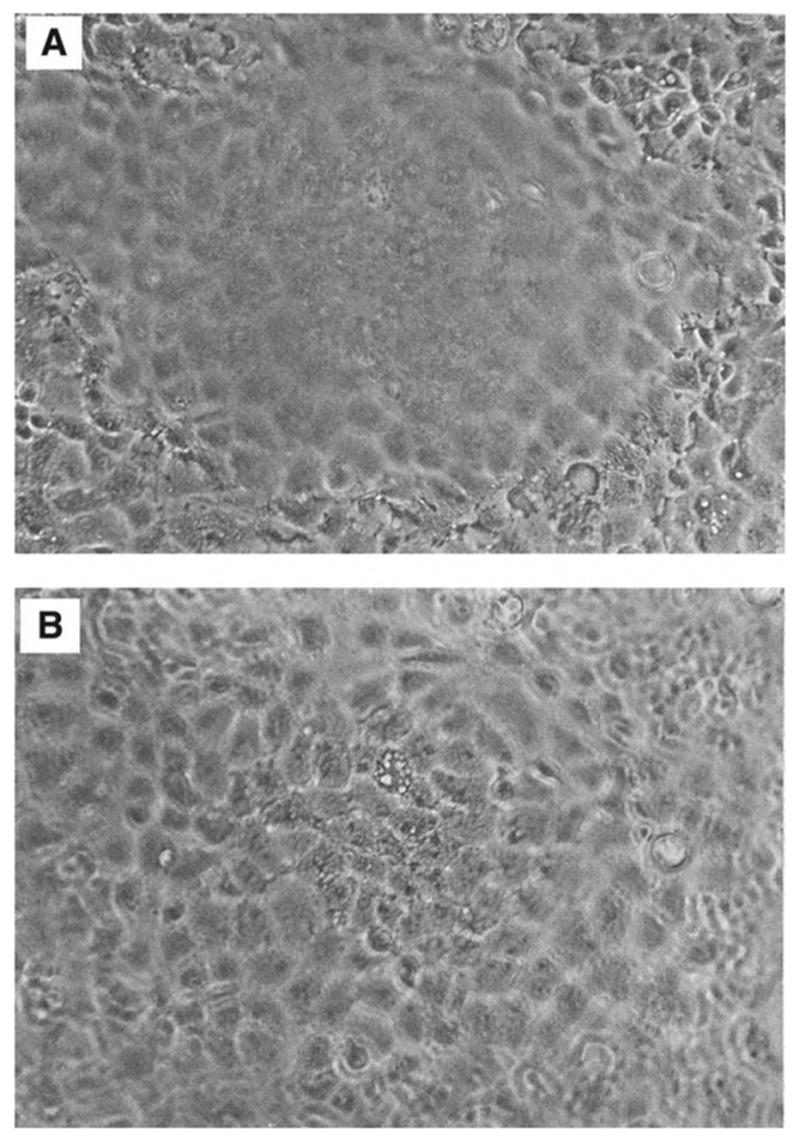
Fig. 1.

Dome formation by rabbit kidney proximal tubule cell cultures. A photomicrograph was taken of a confluent monolayer under an inverted microscope at ×100 magnification. Panel A focuses on the cells in the monolayer, whereas panel B focuses on the cells in the dome.
