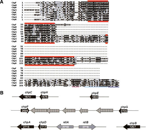
Figure 2.
(A) Alignment of the eight chaplin proteins. The thick red lines mark the chaplin domains. The requirements for covalent cell wall attachment are underlined at the C-terminal end of ChpA–C: the LAXTG sortase recognition motif is underlined in pink, the hydrophobic region following is underlined in light blue, and the positively charged tail is underlined in dark blue. (B) Organization of the chaplin genes. Chaplin genes are shown as black arrows, rodlin genes are shown as gray arrows, and unrelated intervening genes are shown as stippled arrows. Within the rodlin and chaplin gene arrows are the corresponding SCO numbers in white, indicating the position of the gene in the chromosome (genes are numbered 1–7825).

