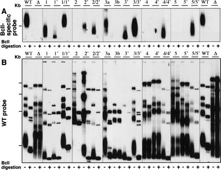
Figure 4.
Base-pairing disruption impairs telomerase action and telomere maintenance in vivo. Southern blotting analysis of the telomeric restriction fragments of the CS3 and CS4 pairing mutants shown in Figure 3B. Digests, wild-type (WT) and Bcl-specific probes, and methods are as described in Figure 2. The P mutants are indicated above each set of lanes. Lanes 1 and 1′ indicate the P1 single mutants, lane 1/1′ indicates the P1 double mutant, and so on. Lanes 3a and 3b are two independent transformant lines of the P3 CS3 mutant (Fig. 3B). For simplicity, in A only the portion of the gel with a group of seven similarly sized telomeric EcoRI restriction fragments (McEachern et al. 2002) in WT K. lactis is shown. All the telomeric restriction fragments are shown in B.

