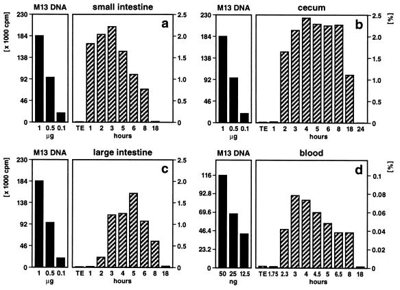
Figure 1.
Quantitation of M13mp18 foreign DNA detectable by dot blot hybridization in different segments of the intestine (a–c) or in peripheral blood (d). Fifty micrograms of M13mp18 DNA (19) (7250 bp) was pipette-fed to 2–6 (12)-month-old mice. As indicated, at times after feeding, the DNA was extracted (1) from the contents of the small intestine (a), the cecum (b), the large intestine (c), or from blood (d). The persisting M13mp18 DNA was identified by dot blot hybridization (1). Aliquot portions of the extracted DNA were fixed on a positively charged Qiagen nylon membrane and hybridized to 32P-labeled (20) M13mp18 DNA followed by autoradiography. In reconstitution experiments, amounts of M13mp18 DNA ranging from 5 ng to 1 μg were also bound to the membranes. Subsequently, autoradiographically localized dots were excised, and the amounts of 32P label membrane bound in these spots were determined by Cerenkov counting. Considering the fraction of the total reextracted DNA from gut segments or blood applied to the dot blot matrix, percentages (scales on right) of the 50 μg of orally administered M13mp18 test DNA were calculated (hatched bars). The scales in cpm and the solid bars on the left referred to the actual 32P Cerenkov cpm for reference M13mp18 DNA.

