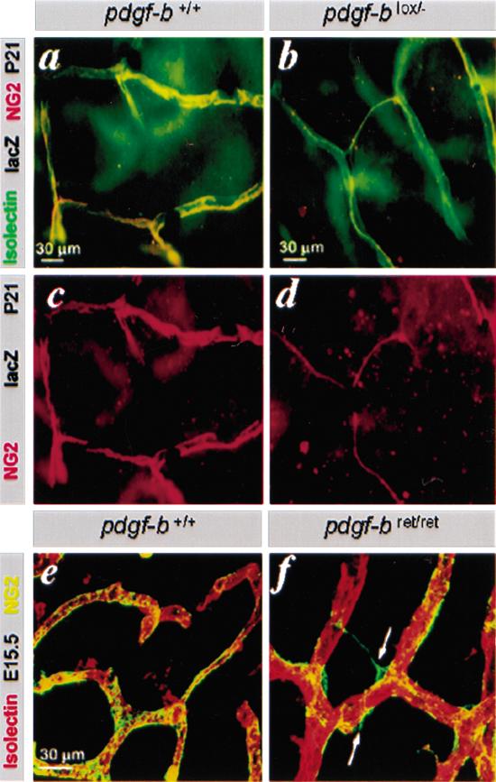
Figure 3.
Defective investment of pericytes into the vessel wall in pdgf-bret/ret mice. (a-d) Isolectin (green), NG2 (red), and LacZ stain of pdgf-b+/+ and pdgf-blox/- P21 retinas. The few pericytes seen in pdgfblox/- mice extend thin endothelium-associated processes. (e,f) NG2 and isolectin staining of E12.5 hindbrain. Pericytes (green) are partially detached and extend processes away from the endothelial cells (red; arrows) in pdgf-bret/ret mice.

