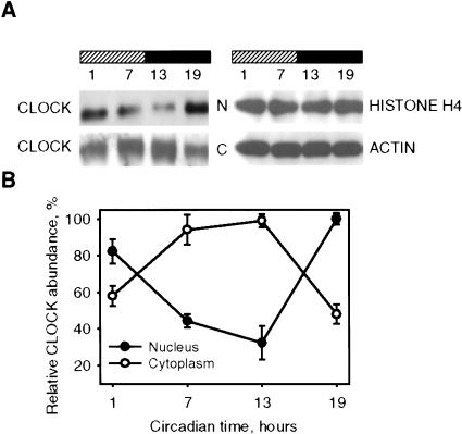
Figure 1.
Circadian oscillation of CLOCK nuclear/cytoplasm distribution in the SCN. (A) SCN extracts obtained from C57BL/6J mice at designated circadian times were fractionated and analyzed by Western blotting using CLOCK antibodies. Histone H4 and β-actin were used for normalization. N, nuclear fraction; C, cytoplasm fraction. (B) Quantitative analysis of CLOCK protein expression in nuclear and cytoplasm fraction of the SCN. Data represent the average of three independent experiments. Each value represents a percentage of maximal level of CLOCK in nucleus or cytoplasm.

