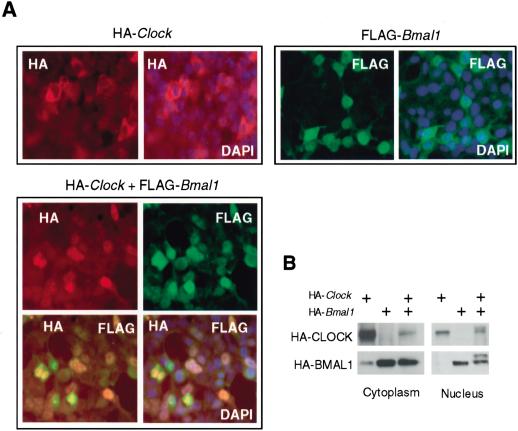
Figure 3.
Nuclear/cytoplasm distribution of ectopically expressed CLOCK and BMAL1. (A) In situ detection of HA-CLOCK and Flag-BMAL1. HEK-293 cells were either individually transfected with pcHA-Clock and pcFlag-Bmal1 or cotransfected with both constructs. Secondary antibodies were conjugated with Texas Red (red fluorescence) for HA-CLOCK detection and FITC (green fluorescence) for Flag-BMAL1 detection. Orange color represents costaining of CLOCK and BMAL1 in cotransfected cells. DAPI (blue fluorescence) staining was used to visualize nuclei. (B) Western blot analysis of nuclear and cytoplasm fractions of HEK-293 cells transfected with pcHA-Clock and pcHA-Bmal1 in different combinations and probed with anti-HA antibodies.

