
Figure 2.
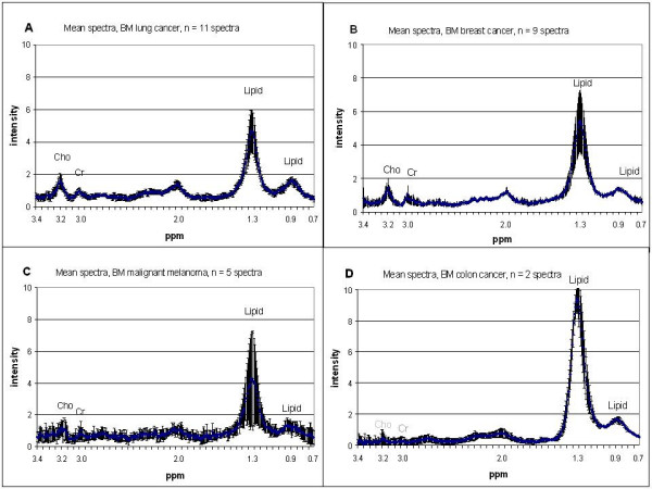
Mean spectra. Short echo time mean spectra ± 95% CI of brain metastases from different primary cancer. A: lung cancer (n = 11 spectra), B: breast cancer (n = 9 spectra), C: malignant melanoma (n = 5 spectra), D: colon cancer (n = 2 spectra). The 0.7 – 3.4 ppm area of the spectra and the detected metabolites (in ppm) are given; tCho (3.2), Cr (3.0): creatine and lipids (1.3, 0.9): methylene and methyl groups.