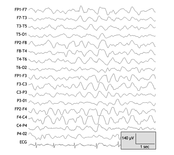
Figure 1: Electroencephalogram taken on day 1 after the patient was admitted to hospital for the treatment of valproate-related hyperammonemic encephalopathy. Note the diffuse high-amplitude generalized slowing, consistent with diffuse cerebral dysfunction. Note: C = central, F = frontal, FP = frontpolar, O = occipital, T = temporal. Odd and even numbers in the electrode locations refer to the left and right hemispheres respectively.
