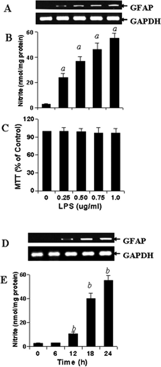
Figure 1.

Dose- and time-dependent production of NO and increase in GFAP expression by LPS in mouse primary astrocytes. Cells were stimulated with different concentrations of LPS under serum-free condition. A–C, After 24 h of stimulation, the expression of GFAP was analyzed in cells by semiquantitative RT-PCR (A), the concentration of nitrite was measured in supernatants (B) by Griess reagent, and the viability was examined by MTT assay (C) as described in Materials and Methods. Results are mean ± SD of three different experiments. ap < 0.001 versus control (LPS, 0 μg/ml). D, E, Subsequently, cells were stimulated with 1.0 μg/ml LPS under serum-free condition for a different time period followed by analysis of GFAP mRNA expression in cells (D) and assay of nitrite in supernatants (E). Results are mean ± SD of three different experiments. bp < 0.001 versus 0 h.
