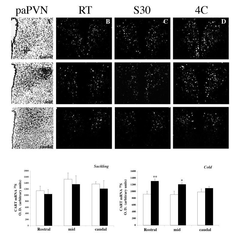
Figure 3.
Distribution of CART mRNA in the parvocellular PVN of lactating rats exposed to 30 min of suckling (column C) or 1h of cold exposure at 4C (column D). CART mRNA was detected by in situ hybridization using a 35S -labeled probe. Note the increase in the density of silver grains in cold exposed animals in the rostral (1st row) and mid (2nd row) sections of the parvocellular PVN (paPVN). First column (A): bright-field illumination showing representative hematoxilin-eosin stained sections trough the rostro-caudal paPVN. Second column (B): CART mRNA pattern in control RT lactating rats; third (C), in lactating rats suckled for 30 min (S30) and last (D) on lactating rats exposed 1h to cold, without pups. Graphs show semiquantitative analysis of CART mRNA after 30’ of suckling or 1hr of cold exposure. Values represent mean ± sem from 4, 8 or 4 slices/animal/group in the rostral, mid and caudal paPVN respectively. (* P < 0.05 or ** P < 0.01 statistically different from control RT animals; Newman-Keuls post hoc testing)

