
Figure 2.
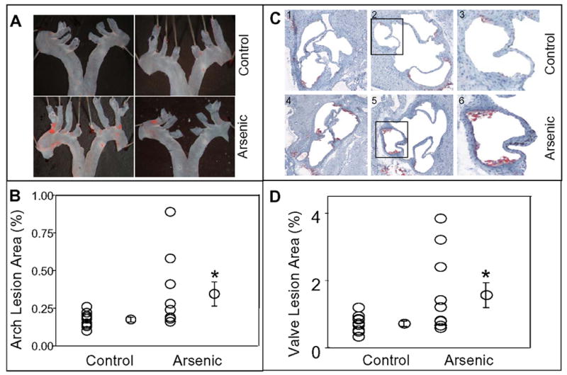
In utero arsenic exposure accelerated atherogenesis in 10 week old ApoE−/− mice. Panel A shows representative photographs of Sudan IV staining in the aortic arch en face of mice exposed (Arsenic) and unexposed (Control) to arsenic in utero. Quantitation of lesions as per cent of aortic arch surface area is illustrated in panel B. Panel C (frames 1 – 6) shows the representative photomicrographs (10x) of aortic valves of mice exposed to arsenic in utero (frames 1 – 3) and unexposed (frames 4 – 6). Lipids were visualized with oil red O staining. Frames 3 and 6 show higher magnification image of the marked sections of images 2 and 4 respectively. Panel D, shows quantitation of lesions as per cent of aortic valve area. Values are expressed as Mean ± SE; * indicates p<0.05.