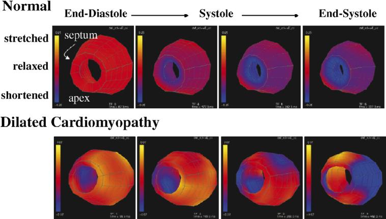
Fig. 3.

These colorized surfaces show the circumferential stretch (yellow) and contraction (blue) of the midwall of a normal human left ventricle (top) and a left ventricle of a patient with DCM (bottom). The apex is toward the viewer and the free wall is on the right side. Four time frames are shown from the beginning of systole through end-systole. Note the early contraction of the septum and late contraction of the free wall in the patient.
