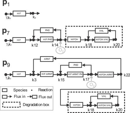
Figure 5. Graphical Representations of the Three Underlying Pathways of HIFα Degradation: p 1, p 7, and p 9 .
Through p 1, HIFα is degraded directly by k 2. In p 7, HIFα first binds with PHD after synthesis. HIFα:PHD is then dissociated into PHD and HIFαOH with the participation of oxygen. HIFαOH then binds with VHL to form HIFαOH:VHL. Finally, the dissociation of HIFαOH:VHL concludes HIFα degradation. The pathway p 9 differs from p 7 only in that HIFα first binds with ARNT after synthesization.

