
Figure 1.
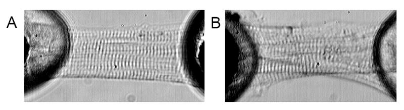
Representative micrographs of cell fragments purified from human cardiac specimens. Panel A shows a cell fragment that was isolated from tissue that had been fast frozen in liquid N2 promptly following surgical excision and generated meaningful F:Ca data. Panel B on the right is a cell fragment that was not preserved as carefully and, despite the intact sarcomere registration, did not generate reliable data. Note the granular deposits and distorted myofibrillar organization.