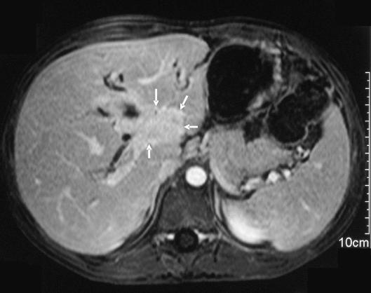
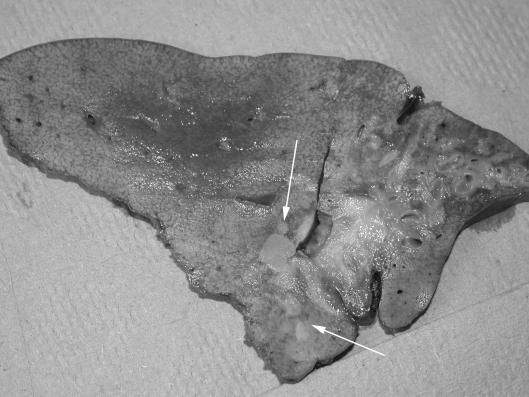
Figure 6.
(A) Axial magnetic resonance image of a 7-year-old boy with an aggressive hilar inflammatory myofibroblastic tumour (arrows). (B) Gross appearance of resection specimen after hepatectomy and OLT. Note the dense hilar infiltration, left lobe atrophy and satellite nodules (arrows).69


