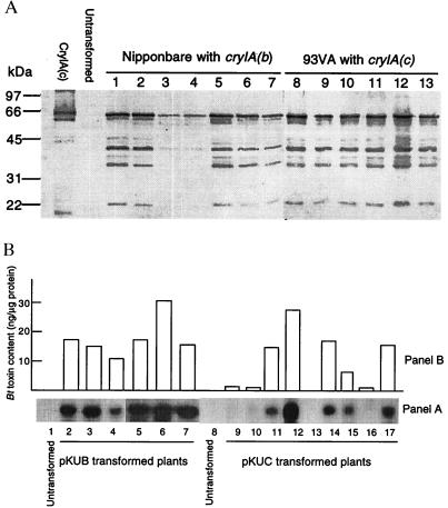
Figure 3.
Expression of cryIA(b) and cryIA(c) in Agrobacterium-transformed rice plants. (A) Western analysis of Bt toxins in transformed rice plants; 2–4 μg of proteins extracted from untransformed and transformed plants were subjected to 10% SDS/PAGE, transferred to nitrocellulose membrane, and reacted with a polyclonal antibody specific to CryIA(b). Samples from two cultivars Nipponbare (transformed with pKUB, lanes 1–7) and 93VA (transformed with pKUC, lanes 8–13) are shown in the blot, together with the CryIA(c) from B. thuringiensis as standard. (B) Comparison of the levels of the cryIA(b) and cryIA(c) transcripts and Bt toxins. (Panel A) Northern blot analysis of cryIA(b) and cryIA(c) transcripts in plants transformed with pKUB (lanes 2–7) and pKUC (lanes 9–17). Total RNA (10 μg/lane) was extracted from leaf tissues, separated electrophoretically on 1.2% agarose-formaldehyde gel, blotted to Hybond-N nylon membrane, and hybridized to a DIG-labeled cryIA(b) fragment. (Panel B) Corresponding Bt toxin levels in the plants used for Northern analysis. The Bt toxin levels were determined by comparison of intensities of the immunologically developed color from the plant samples with that from the purified CryIA(c).

