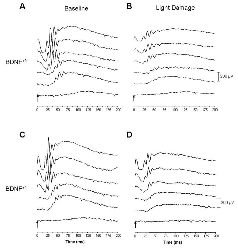
Figure 2.

Families of ERGs elicited from BDNF+/+ (A, C) and BDNF+/− (B, D) mice before (A, B) and after (C, D) light damage, at increasing light intensities (40, 30, 20, 10, 6, and 0 log units of attenuation). At higher light intensities, rod ERG amplitudes were approximately 25% smaller in BDNF+/− mice than in their age-matched wild-type littermates. Light damage resulted in a large reduction of a- and b-wave amplitudes in the BDNF+/+ mouse retina (maximum amplitudes were reduced by approximately 40% and 60%, respectively) but had a less deleterious effect on the ERG elicited from the BDNF+/− retina (maximum amplitudes were reduced by only approximately 13% and 40%, respectively). For complete data analysis, see Figure 3, Table 2, and Results section.
