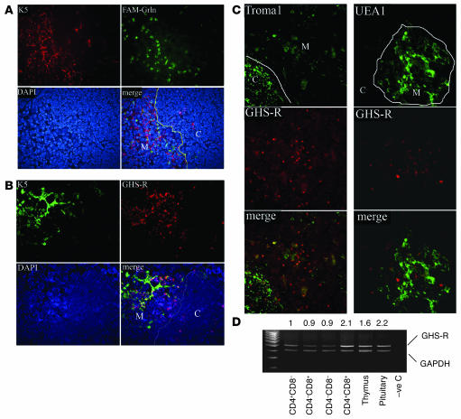
Figure 1. The GHS-R expression in thymus.
(A) The ghrelin binding sites exist largely in the medullary area of the thymus. Frozen thymic sections were labeled with FAM-conjugated ghrelin (FAM-Grln) and stained with medullary epithelial cell marker, Alexa Fluor 594–conjugated anti–Ker5 (K5) antibody and counter-stained with DAPI for nuclei. Ghrelin binding sites (green) are largely segregated from Ker5-expressing cells in the cortex (C; red), as shown by lack of a yellow staining pattern, which would suggest colocalization (line depicts the CMJ in the thymus). M, medulla. (B) Similar to FAM-ghrelin binding, the GHS-R antibody–positive cells displayed only modest colocalization with Ker5+ medullary TECs. (C) GHS-R (red) displayed little to no colocalization with cortical Ker8+ and medullary UEA-1+ TECs. However, a minor subset of Ker8+ cells in the medulla displayed GHS-R immunopositivity. (D) Thymus and sorted SP, DP, and DN thymocytes express GHS-R mRNA. DP cells expressed 2-fold higher GHS-R than did SP and DN cells, with levels comparable to those of the pituitary. Numbers above the blot indicate the ratio of GHS-R to GAPDH expression. –ve C, negative control. Original magnification, ×10.

