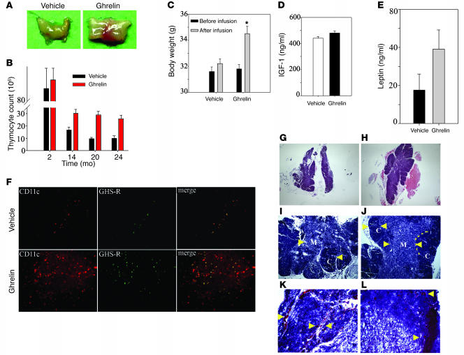
Figure 3. Ghrelin enhances thymic size and cellularity in aging mice.
(A) Ghrelin infusion for 2 weeks via s.c. osmotic pumps caused a significant increase in thymic size in 14-month-old animals. (B) Ghrelin did not alter thymocyte counts in the 2-month-old mice but significantly increased total thymocyte numbers in 14-, 20- and 24-month-old mice. (C–E) Ghrelin treatment significantly increased body weight in 14-month-old mice (C) without a significant change the peripheral IGF-1 (D) or leptin (E) levels. *P < 0.05. (F) Ghrelin infusion into 14-month-old mice led to increased GHS-R expression (green) with partial colocalization with CD11c+ cells. In addition, ghrelin administration resulted in an enhanced number of CD11c+ dendritic cells in the thymic medulla. (G–J) Compared with 14-month-old vehicle-infused animals, ghrelin infusion significantly improved the thymic architecture. Ghrelin infusion is associated with increased cellularity, clear delineation (arrowheads) of cortex (dark stain) from medulla (light stain) and well defined CMJ (dotted line), the site where progenitors arrive in thymus. (K and L) Frozen thymi from 14-month-old mice stained for Oil Red O displayed an increased number of “adipocyte-like” lipid-laden cells (arrowheads) in the parenchyma and perivascular space, while in ghrelin-infused mice, a marked reduction in PVS (K and L) with reduced numbers of lipid droplet–containing cells (L). Original magnification, ×5 (G and H); ×10 (F, I, and J); ×20 (K and L).

