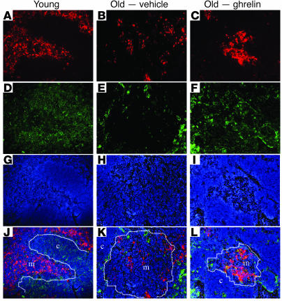
Figure 4. Ghrelin renews the TEC compartment during aging.
The thymus from young mice displays abundant UEA-1+ cells (A–C; red) and Ker8+ cells (D–F; green) in the medulla and cortex. (B, E, H, and K) The aging thymus of 14-month-old mice displayed diffuse and reduced medullary UEA-1+ cells and Ker8+ cortical TECs. (C, F, I, and L) However, compared with vehicle-treated aged mice, ghrelin infusion resulted in a significant increase in both medullary and cortical TECs based on the increase in UEA-1 and Ker8 staining. (G–I) DAPI staining to visualize the nuclei of the cells. (J–L) Combined images of the upper panels. Yellow staining denotes colocalization. Original magnification, ×10.

