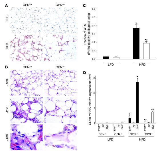
Figure 4. OPN deficiency decreases ATM content in obese mice.
(A) ATM content was determined by immunohistochemical analysis of epididymal adipose tissues isolated from OPN+/+ and OPN–/– mice fed a LFD or HFD. Adipose tissues were stained using an absorbed rabbit anti-mouse macrophage antiserum (original magnification, ×100). (B) Epididymal adipose tissues from obese OPN+/+ and OPN–/– mice were analyzed for macrophage content using an F4/80 antibody (magnified as indicated). (C) Macrophage content was quantified by analyzing the fraction of F4/80-stained cells relative to total number of cells in epididymal adipose tissue from OPN+/+ (black bars) and OPN–/– (white bars) mice fed a LFD or HFD (n = 8/group). Values are expressed as mean ± SEM. (D) Macrophage content was quantitatively assessed by real-time RT-PCR for CD68 mRNA expression in EWAT, the AF, and the SVF isolated from OPN+/+ (black bars) and OPN–/– (white bars) mice (n = 12/group) fed a LFD or HFD for 25 weeks. Data are presented as relative CD68 mRNA expression normalized to TFIIB mRNA expression and are expressed as mean ± SEM. *P < 0.05, HFD compared with LFD; #P < 0.05, OPN–/– mice compared with OPN+/+ mice fed HFD.

