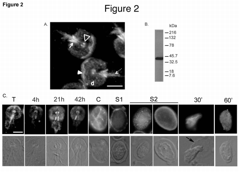
Fig. 2.

A. Immunolocalization of gPP2A‐C to the Giardia cytoskeleton. Cytoskeletons of vegetative trophozoites were probed with the anti‐PP2A‐C antibody. The filled arrowhead shows the anterior PDR, the open arrowhead shows the basal bodies. The solid arrow points to posterior‐lateral PDR and the dashed arrow to the caudal PDR. “d” is the ventral disk. Bar = 5 µm
B. Antibody specificity: Western blot of a total Giardia lysate reacted with the anti‐PP2A‐C antibody used for immunolocalization.
C. Immunolocalization of gPP2A‐C changes during Giardia differentiation. Cytoskeletons of vegetative (T), 4 h, 21 h and 42 h encysting trophozoites, whole water‐treated cyst (C), excysting cysts in Stages 1 (S1) and 2 (S2) and 30 (30’) and 60 (60’) min emerging cells were probed with the same anti‐PP2A‐C antibody. Note: Intensities of the signals in the panels should not be compared as the exposure times were optimized individually. The black arrow points to the degraded cyst wall surrounding an emerging excyzoite. Bar = 5 µm
