
Fig. 3.
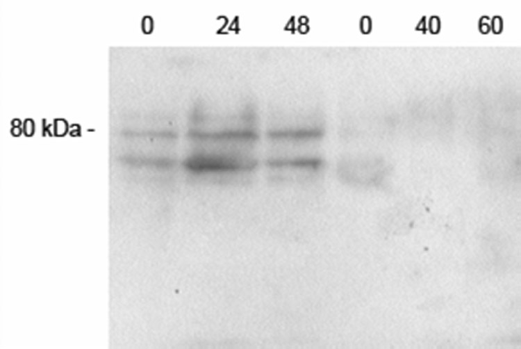
Immunoblotting of cultured erythroid cell membranes for the SVCT2. The cells were rinsed once in PBS and were extracted with lysis buffer. Protein from approximately 5 × 10⁷ cells was applied to each lane of the gel. Electrophoresis and staining for SVCT2 was carried out as described under Methods. The location of the relevant molecular weight marker is shown to the left, and each lane is identified by the hours in culture before extrusion of the nucleus at 48 h and after extrusion to 60 h.