
Fig. 4.
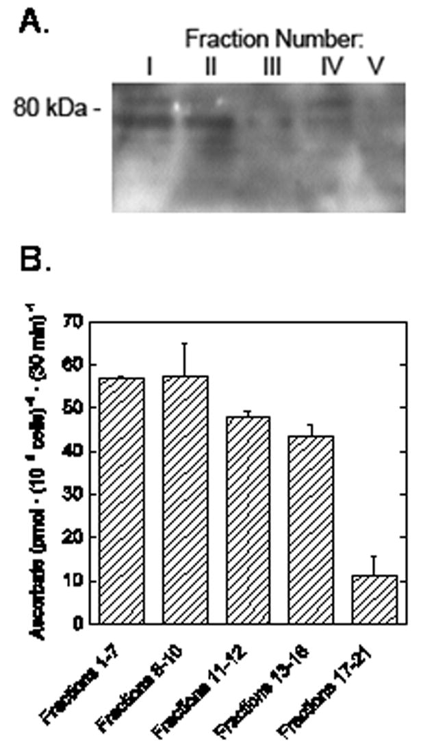
SVCT2 content and ascorbate transport in various erythroid fractions. Sedimentation velocity was used to separate cells cultured for 44 h into 5 fractions as described in the text. The cells in each fraction were rinsed in PBS and extracted with lysis buffer. Panel A. Membranes from approximately 5 × 10⁷ cells were loaded into each gel lane. Electrophoresis and immunoblotting were carried out as described under Methods. The location of the relevant molecular weight marker is noted on the left. Panel B. Ascorbate transport was measured in duplicate in cells after fractionation. Results are shown from one of two such experiments performed.