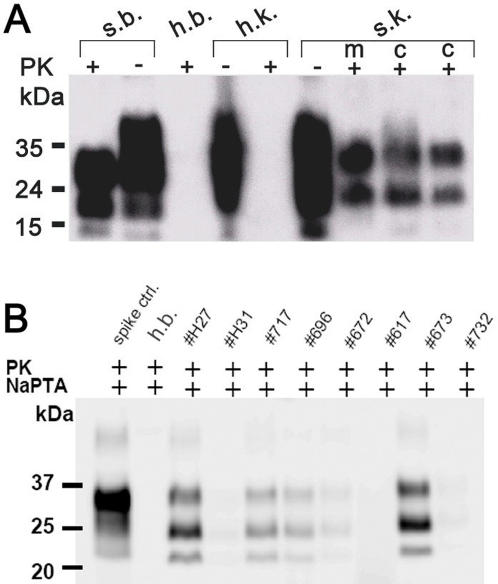
Figure 1. Western Blotting PrPSc deposition in scrapie-affected Sarda sheep.
(A) Detection of PrPSc by conventional Western blot in the cortical and medullar part of kidneys. Brain and kidney homogenates were treated with (+) or without (−) proteinase K (PK), amounts of kidney and brain represented in each line are 1mg and 1g respectively. S.b. = brain derived from a naturally scrapie-sick sheep; h.b. = brain derived from a scrapie-free sheep control; h.k. = kidney derived from a scrapie-free sheep control, s.k. = kidney derived from a naturally scrapie-sick sheep. kD = kilo Dalton, M = medulla, C = cortex. (B) Detection of PrPSc by NaPTA Western blot in kidney homogenate derived from different Sarda sheep. Substantial individual variation of the Western blotting signal was observed among the sheep examined. Spike control = brain homogenate derived from a scrapie–sick sheep spiked into negative control kidney homogenate. h.b. = brain derived from a scrapie-free sheep control. 2000–4000 µg of renal homogenate were used as a starting material to perform NaPTA precipitation.

