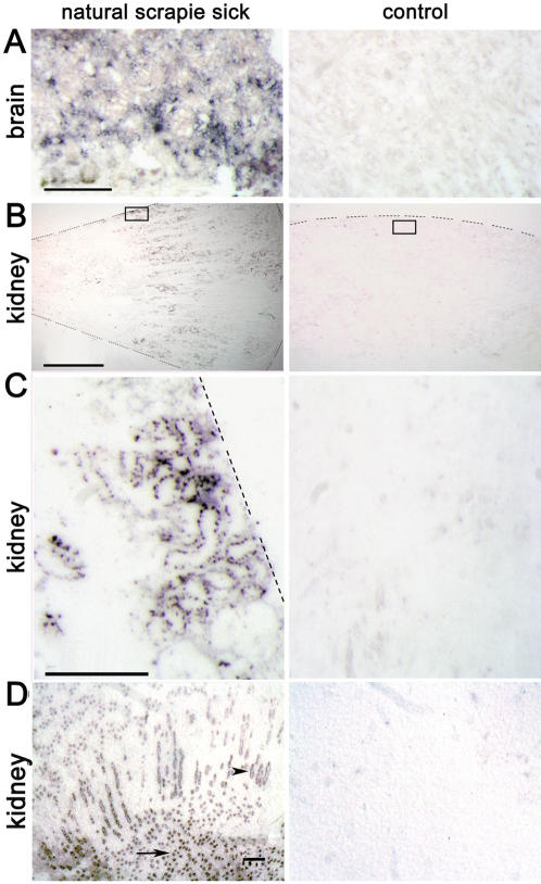
Figure 2. PET blot analysis identifying PrPSc in the tubular structures of the cortex and medulla in naturally scrapie-sick Sarda sheep.
(A) Brains of scrapie-sick (left) and healthy sheep (right) as a control for PET blot analysis procedure. Scale bar = 100 µm. (B) Low magnification of transverse paraffin sections from kidneys of a naturally scrapie-sick and a healthy sheep demonstrating PrPSc in the medullary and cortical regions of the kidney. Dotted lines indicate the locus of the kidney capsule or the edge of the tissue section. The small transparent box indicates the region shown at higher magnification and resolution in figure C. Scale bar = 1 cm. (C) PrPSc deposits and tubular structure fit together in the cortical part of the kidney in scrapie-sick sheep. Scale bar = 100 µm. (D) PrPSc is also found to a high degree in the collecting ducts of the medulla (arrowhead) as well as in the papillae (arrow) of kidneys derived from naturally scrapie infected sheep. Scale bar = 30 µm.

