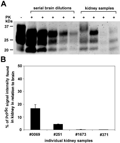
Figure 4. Quantification of PrPSc in renal homogenates of naturally scrapie-sick sheep.
(A) Conventional Western blot analysis of various brain dilutions after proteinase K (PK) digestion. Undigested brain is loaded to control for PK digest (lane 1). Kidney homogenates from independent scrapie sick animals were loaded (lanes 7–10). (B) Relative quantification of PrPSc signal intensity found in kidney homogenates in relation to brain derived PrPSc. Our results indicate that the highest amounts of renal PrPSc detected were appr. up to 20% of the PrPSc signal detected in brain.

