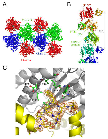
Figure 1. Crystal packing and quaternary structures.
(A) SsoRadA protomers packed into three extended helical filaments. Chain A was located at the origin of the unit cell, whereas chains B and C were located one-third and two-third diagonal to the unit cell. (B) Side view of the SsoRadA right-handed helical filament crystal structure. The helical pitch of the filament is 98 Å. Each protomer is shown in a different color. The N-terminal domain (NTD), polymerization motif (PM), and central ATPase domain are indicated. (C) The Phe73 of the PM is buried in the hydrophobic pocket of the neighboring ATPase domain. Several hydrophobic residues that interact with the Phe73 side chain are indicated. The interactions result in the assembly of SsoRadA protomers into a filament. 2F o–F c electron density maps (contoured at 1.0 σ), corresponding to the PM are shown in orange.

