Figure 1.
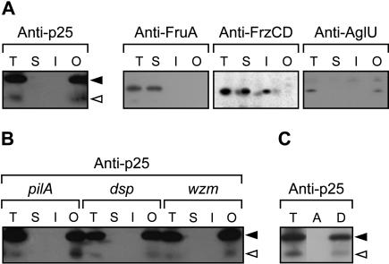
Biochemical fractionation of starved M. xanthus cells. (A) Wild-type cells (DK1622) were starved for 24 h on TPM-agar. The total cell lysate (T) was separated into fractions enriched for soluble proteins (S), inner membrane proteins (I), and outer membrane proteins (O). The fractions were subjected to immunoblot analyses and probed with anti-p25 antibodies. As controls, the association of FruA, FrzCD, and AglU with the different fractions was determined using anti-FruA, anti-FrzCD, and anti-AglU antibodies, respectively. (B) pilA cells (DK10407), which are unable to synthesize type IV pili; dsp cells (DK3470), which are unable to synthesize extracellular fibrils; and wzm cells (HK1321), which are deficient in lipopolysaccharide O-antigen synthesis, were starved and fractionated as in A and probed with anti-p25 antibodies. (C) DK1622/pTK98-10 cells were starved for 24 h. The lysate was phase separated with Triton X-114 into an aqueous (A) and a detergent (D) phase. The immunoblots were probed with anti-p25 antibody. Closed and open arrowheads indicate p25 and p17, respectively.

