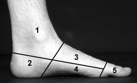
Figure 1.
The zones of the foot used to analyse the data as described by Kirby et al.2 The lines correspond to an oblique coronal plane, drawn from the mid-tarsal joint to the posterior margin of the longitudinal arch; a transverse plane, drawn from the mid-point of the metatarsal heads to the level of insertion of the Achilles tendon into the calcaneus; and a coronal plane, drawn through the metatarsophalangeal joints. These regions were numbered 1–5, to correspond to the ankle, heel, dorsum of the foot, plantar surface of the foot, and toes.

