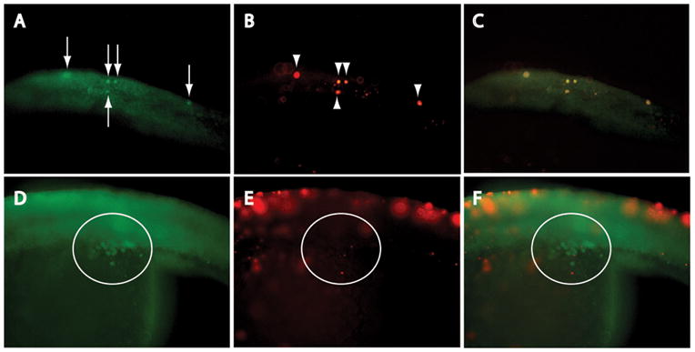
Figure 5.

Ectopic PGCs undergo apoptosis. (A) Vasa immunostaining, and (B) TUNEL analysis of ectopic PGCs in dorsal trunk of igf1rb-MO-injected embryo; (C; merged image) colocalization of Vasa-positive (arrows) and TUNEL-positive (arrowheads) cells confirms DNA fragmentation in ectopic PGCs. (D) Vasa-positive, but (E) TUNEL-negative PGCs in genital ridge (circled) of igf1rb-MO-injected embryo; images merged in (F). An absence of DNA fragmentation in genital ridge PGCs confirms survival of the subpopulation of PGCs that successfully migrate to the genital ridge. Anterior is to the left in all images; magnification 200x.
