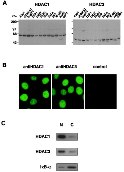
Figure 2.
HDAC1p and HDAC3p are ubiquitously expressed and predominantly nuclear proteins. (A) Western blot analysis. Western blots containing 20 μg of protein per lane from different cell lines were developed with a polyclonal serum raised against HDAC1p or HDAC3p. Coomassie blue staining of gels showed that similar amounts of cellular protein were loaded in different lanes (data not shown). Sizes of protein markers are indicated. (B) Immunofluorescence. HeLa cells grown on coverslips were fixed, stained with the anti-HDAC1 or anti-HDAC3 antiserum, and examined by immunofluorescence microscopy as described (20). (C) Subcellular localization of HDAC1p and HDAC3p. Jurkat cells nuclear (N) and cytoplasmic (C) fractions were prepared. Proportional amounts of each fraction were analyzed by Western blotting using specific antiserum directed against HDAC3, HDAC1, or IκB-α.

