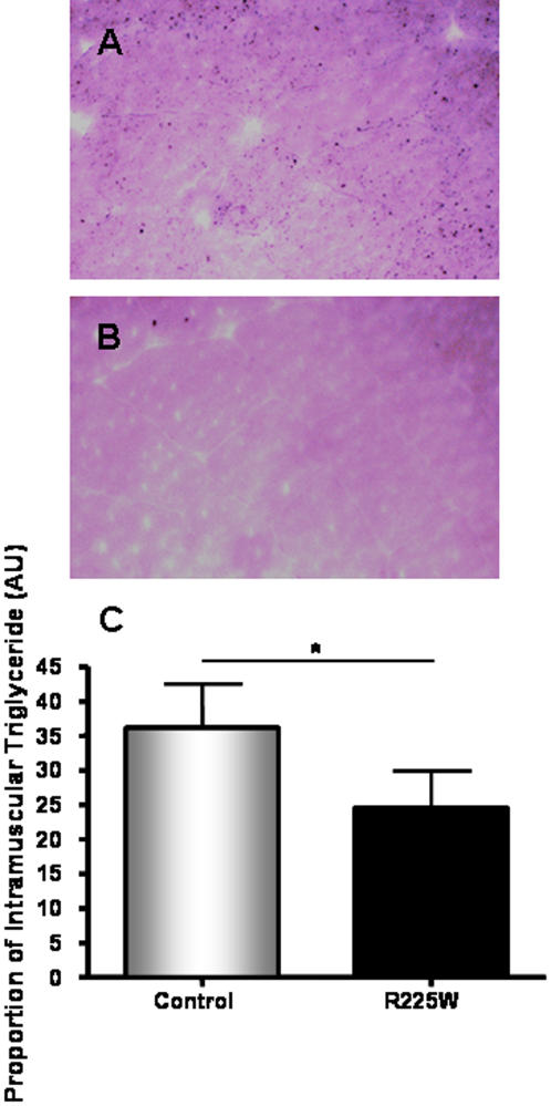
Figure 6. Intramuscular triglyceride content of vastus lateralis for R225W and control subjects.
Muscle sections were prepared and stained with OsO4, as described in the methods section (A: control, B: R225W). C: quantification of IMTG via imaging software. Means +/− SD. n = 4, Student's t-test, *p = 0.032.

