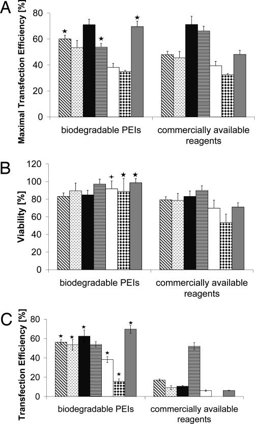
Fig. 6.
Comparison of biodegradable PEIs with commercially available transfection reagents. (A and B) EGFP-positive cells expressed as maximal transfection efficiency (A) and corresponding cell viability (B) of various biodegradable PEIs and commercially available transfection reagents complexed with pEGFP-N1 in (from left to right) CHO-K1 (diagonally hatched bars), COS-7 (horizontally hatched bars), NIH/3T3 (black bars), HepG2 (horizontally striped bars), HCT116 (white bars), HeLa (diamond-hatched bars), and HEK-293 (gray bars) cells as determined by flow cytometry. (C) Transfection efficiency under conditions where cell viability is >90%. Statistically significant differences of biodegradable PEIs compared with other transfection reagents are denoted by  (P < 0.05) or by ★ (P < 0.01).

