Fig. 3.
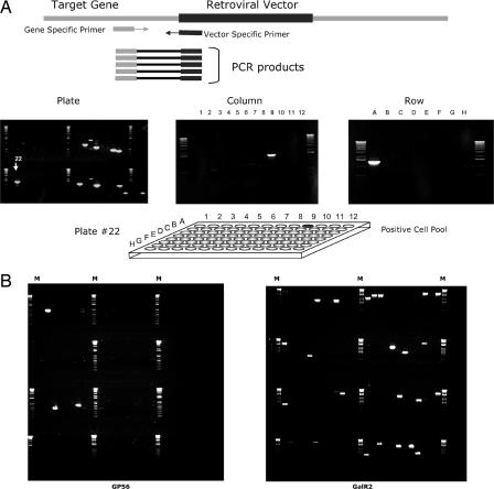
Library screening. (A) Nested PCR with gene- and vector-specific primers (Top) was used to screen plate pools from multiple library units (Middle Left). One lane corresponds to one plate pool. The example shows screening of four units, with 10 plate pools in each unit for a gene of interest. Multiple insertions (PCR bands) were detected in different plate pools. Then column and row pools (Middle Center and Right, respectively) of the unit(s) that demonstrated the presence of a sequencing-confirmed insertion in the gene were screened by using the same pairs of primers to find the 3D address of the positive well in the library (e.g., the positive well shown at Bottom gave rise to identical PCR fragments in plate pool no. 22, column pool no. 9, row pool A). (B) The results of screening eight library units (≈4 × 10⁶ mutant ES cell clones) for insertions in the genes of two different GPCRs. M, marker.

