Fig. 5.
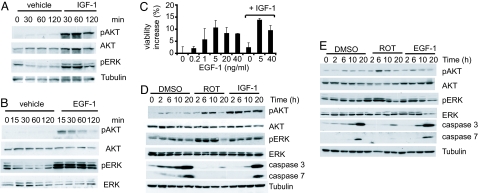
Growth factors activate ERK and AKT and rescue cell death. ERK and AKT activations after treatment with IGF-1 (90 ng/ml) (A) and EGF-1 (5 ng/ml) (B) in Sdm were assayed by Western blotting. (C) N548 mutant cells were treated with EGF-1 or IGF-1 alone or in combination, and cell viability was assayed after 2 days in Sdm by trypan blue dye exclusion assay. The results are the averages ± SD of one representative experiment. IGF-1 was used at one concentration (90 ng/ml); lower concentrations were ineffective, and higher concentrations did not further enhance viability. (D and E) N548 mutant cells were treated with DMSO, rotenone (Rot), or IGF-1 (90 ng/ml) (D) or EGF-1 (5 ng/ml) (E) in Sdm, and activation of AKT, ERK, and cleaved caspase 3 and 7 levels were monitored by Western blotting. Tubulin served as a loading control.

