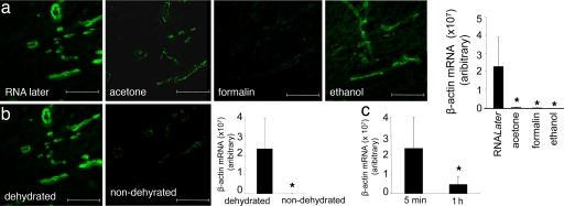
Fig. 2.
Optimization of vessel staining protocol: fixation and dehydration. (a) Human wound sections were subjected to standard fixation methods, i.e., RNALater, acetone, formalin (neutral buffered formalin), or ethanol (95% vol/vol ethanol) as shown. After fixation, sections were stained with UEA I lectin (green). The bar graph shows relative β-actin mRNA levels quantified by using real-time PCR. RNA was extracted from 400,000 μm2 of vessel elements captured after laser microdissection after specified fixation and UEA I lectin staining. *, P < 0.05 lower compared with the RNALater treated group. (b) UEA I stained dehydrated versus nondehydrated wound tissue sections. The bar graph shows relative β-actin mRNA levels quantified from tissue elements captured from RNALater fixed and dehydrated or nondehydrated. *, P < 0.05 lower compared with the dehydrated group. (c) Stability of β-actin transcript in tissue sections as a function of time after RNALater fixation, staining, and dehydration. Vessel area (400,000 μm2) was processed, and β-actin expression was quantified as described. *, P < 0.05 lower compared with 5-min group. (Scale bars: 200 μm.)

