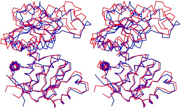
Figure 3.
Stereo view of the Cα backbone of PF (blue) and PA (red) monomers, where the two CP binding domains have been superimposed. The ornithine binding domains differ in a rotation of 8°, resulting in a domain closure in PF OTCase. The CP binding domains are at the bottom. The figure was generated by using molscript (36).

